🩺 DermDepth — Monocular Metric-Scale 3D for Dermatology
Dermatology is largely a measurement problem: clinicians screen and monitor lesions and wounds by tracking size, border, elevation and texture over time. Those properties are inherently 3D — yet point-of-care imaging is almost always a single 2D photo.
DermDepth recovers metric-scale 3D from one ordinary photograph — no depth sensor, no second view, no ruler in frame. A 2.1M-parameter scale-and-normal head sits on a frozen MoGe-2 backbone, trained progressively on D-Synth (synthetic renders with pixel-perfect depth, normals and intrinsics) and then on real clinical data. On the paper's held-out benchmarks it cuts metric scale error from 16.1× to 1.15× on SKINL2 and from 81× to 1.95× on DDI, and reduces Fitzpatrick skin-tone scale disparity from 10.90 to 1.02. Those are benchmark figures — accuracy on your own photograph, from an unfamiliar camera or distance, may be substantially worse.
Reconstruct an image, then use 📏 Measure distance to click two points and read the estimated metric distance between them.
⚠️ Research demonstration only — not a medical device. These outputs are not diagnostic and must not inform clinical decisions. Every distance, area and volume shown is a model estimate from a single photograph, not a measurement — treat them as comparative, never absolute. Predictions on out-of-distribution images can fail silently.
How it works
| Output | Checkpoint | Why |
|---|---|---|
| Metric depth + 3D mesh | DermDepth_Synth_SKINL2_WoundsDB_DDI.pt |
The paper's best model — D-Synth → SKINL2 + WoundsDB → DDI pseudo-GT for metric scale. |
| Surface normals | DermDepth_Synth_Normals.pt |
Normal-head model trained on D-Synth, whose rendered normals are the only clean normal supervision (real ToF/plenoptic normals are noisy). |
📏 Measure distance reports the estimated chord and along-the-surface arc between two points; 📐 Measure volume estimates 3D area and raised/cavity volume for a painted region. Both read through the predicted metric point map, so they are estimates in millimetres rather than pixel counts — not ground truth.
Example credits
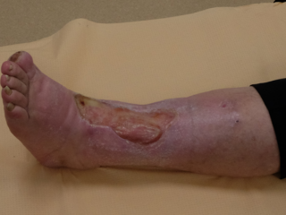
The first three are real clinical photographs from WoundsDB (Chronic Wounds
Multimodal Image Database, Silesian University of Technology), used under
CC BY 4.0 at their native 320×240 — the
resolution the paper evaluates WoundsDB at. They are held-out cases (the paper splits
WoundsDB by case: 1–30 train, 31+ test): case_45 (leg), case_33 (hand), case_42 (foot).
Kręcichwost, M., Czajkowska, J., Wijata, A., Juszczyk, J., Pyciński, B., Biesok, M., Rudzki, M., Majewski, J., Kostecki, J., & Pietka, E. (2021). Chronic wounds multimodal image database. Computerized Medical Imaging and Graphics, 88, 101844. doi:10.1016/j.compmedimag.2020.101844
The last three are synthetic renders from D-Synth (Carrión & Norouzi), CC BY-NC 4.0 — one per Fitzpatrick group (I–II, III–IV, V–VI). They are renders, not patient photographs, and imply no diagnosis.
No DDI imagery is bundled: Stanford's Research Use Agreement prohibits redistributing any portion of that dataset.
Links
📄 Paper (MICCAI 2026) · 🤗 Model · 📊 D-Synth dataset · 💻 Code